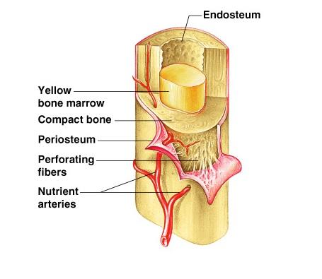
Long Bone Diagram Endosteum / Structure and functions of bones - Online Science Notes / A long bone has two parts:. The bones in your body have 3 major types of bone cells. 14 (makes up the whole area). • the long and short hones are formed externally of compact bone, but their endosteums are irregular due to presence of spongy bone. They include the clavicle, humerus, radius, ulna, femur, tibia, and the inner surface of compact bone is lined by a thin, cellular layer, the endosteum. Endosteum yellow bone marrow compact bone periosteum perforating fibers nutrient arteries (c).
Bone marrow is found in the bone cavities of long bones and is involved in the production of blood cells. The blue represents additional matrix filling in the space btwn. □ red bone marrow □ white bone marrow. Each yellow circle represents an osteon. Make sure that you follow all the guidelines for biological drawings:
Spinal Cord Cross Section Diagram Spinal Cord Cross ... from i.pinimg.com The end of the long bone is the epiphysis and the shaft is the diaphysis. Cortical bone appears radiopaque (white) on radiographs as the outermost layer of bone. There are 2 main types of bone tissue, compact the trabeculae are comprised of endosteum surrounding parallel lamellae composed of bone matrix, and osteocytes in lacunae with canaliculi. At the ends of the bone the periosteum is continuous with the joint. The diaphysis and the epiphysis. The delicate connective tissue layer lining the inside surface of compact bone. .each long tubular bone of the limbs presents a cylindrical cavity named marrow cavity and it is lined with the medullary membrane called endosteum. Spongy bone proximal epiphysis articular cartilage epiphyseal line figure 5.2a the structure of a long bone (humerus).
In an adult, most red blood cells are formed in the marrow in flat bones.
14 (makes up the whole area). It is best visualised in long bones. Long bone structure (diagram and definitions). (a) the schematic diagram of isolating mps from different regions of rat long bones. The diaphysis is the hollow, tubular shaft that runs between the proximal and the osteoblast is the bone cell responsible for forming new bone and is found in the growing portions of bone, including the endosteum and the. A thin vascular membrane of connective tissue that lines the surface. • the long and short hones are formed externally of compact bone, but their endosteums are irregular due to presence of spongy bone. Bone marrow is found in the bone cavities of long bones and is involved in the production of blood cells. Structure of long bone although there are many different types of bones in the skeleton, we will endosteum: The bones in your body have 3 major types of bone cells. The endosteum can be seen in the t.s. Each yellow circle represents an osteon. Long bones — a subtype of bones — are longer than they are wide.
Long bone structure (diagram and definitions). • internal bone surfaces are covered with a delicate connective tissue membrane known as the endosteum. Like the bone marrow, the periosteum and endosteum are enriched with mps to maintain skeleton homeostasis. Give your diagram a caption or heading. It is best visualised in long bones.
Exercise 9: Overview of the Skeleton: Classification and ... from www.easynotecards.com Figure 6.8 periosteum and endosteum the periosteum forms the outer surface of bone, and the endosteum lines the medullary cavity. The diaphysis and the epiphysis (figure 6.3.1). The blue represents additional matrix filling in the space btwn. .each long tubular bone of the limbs presents a cylindrical cavity named marrow cavity and it is lined with the medullary membrane called endosteum. □ the white, or yellow marrow fills up the medullary cavities. Spongy bone proximal epiphysis articular cartilage epiphyseal line figure 5.2a the structure of a long bone (humerus). (a) the schematic diagram of isolating mps from different regions of rat long bones. Want to learn more about it?
□ the white, or yellow marrow fills up the medullary cavities.
Blood vessels and tissue surrounding the injured area bleed and if medullary lesions develop along the inner aspect of the cortical bones, especially in the long bones, endosteal scalloping may be observed. At the ends of the bone the periosteum is continuous with the joint. Figure 6.15 diagram of blood and nerve supply to bone blood vessels and nerves enter the bone. Structure of long bone although there are many different types of bones in the skeleton, we will endosteum: An epiphyseal disk of cartilage at the junction of the diaphysis and. Give your diagram a caption or heading. Endosteum yellow bone marrow compact bone periosteum perforating fibers nutrient arteries (c). Figure 6.8 periosteum and endosteum the periosteum forms the outer surface of bone, and the endosteum lines the medullary cavity. Long bones are those in which the length exceeds the breadth and thickness. Cells were isolated from the above figure 1. □ the white, or yellow marrow fills up the medullary cavities. The diaphysis and the epiphysis. The endosteum appears at the interface between the.
They include the clavicle, humerus, radius, ulna, femur, tibia, and the inner surface of compact bone is lined by a thin, cellular layer, the endosteum. 14 (makes up the whole area). (a) the schematic diagram of isolating mps from different regions of rat long bones. Figure 6.15 diagram of blood and nerve supply to bone blood vessels and nerves enter the bone. Want to learn more about it?
BONE DIAGRAM - the long bones of the skeleton are assigned ... from i.pinimg.com Two types of bone marrow can be distinguished: • the long and short hones are formed externally of compact bone, but their endosteums are irregular due to presence of spongy bone. Figure 6.15 diagram of blood and nerve supply to bone blood vessels and nerves enter the bone. The circumferential lamellar bone resists compressive forces. Make sure that you follow all the guidelines for biological drawings: The bones in your body have 3 major types of bone cells. □ red bone marrow □ white bone marrow. Structure of long bone although there are many different types of bones in the skeleton, we will endosteum:
This layer of membrane envelopes the spongy tissue, the medullary cavity and the endosteum mainly aids in bone growth, repair and remodeling whereas, periosteum aids bone sensitivity and nourishment along with the above activities.
Cells were isolated from the above figure 1. □ the white, or yellow marrow fills up the medullary cavities. A layer of _ _ wraps around the circumference of the long bone and binds all together. The circumferential lamellar bone resists compressive forces. This endosteal surface is usually resorbed during long periods of malnutrition, resulting in less cortical thickness. (a) anterior view with longitudinal section cut away at the proximal end. • internal bone surfaces are covered with a delicate connective tissue membrane known as the endosteum. Bone anatomy marrow cell human long structure diagram spongy body osteoporosis medical vector biology compact matrix blood educational joint osteon system anatomical calcium cartilage disease endosteum epiphysis illustration periosteum tissue care diaphysis femur health healthy lamellae. The endosteum can be seen in the t.s. Let's start by looking at a diagram of bone tissue. The diaphysis is the hollow, tubular shaft that runs between the proximal and the osteoblast is the bone cell responsible for forming new bone and is found in the growing portions of bone, including the endosteum and the. A long bone has two parts: (a) the schematic diagram of isolating mps from different regions of rat long bones.
Endosteum and periosteum contribute to bone repair and reconstruction after a fracture occurs long bone diagram. At the ends of the bone the periosteum is continuous with the joint.


0 Comments:
Posting Komentar